A Dream Come True
A Scientific Journey by Ben Allweiss
“You’ve got to have a dream, if you don’t have a dream, how you expect to have a dream come true…” From the musical South Pacific
“…lose your dreams and you will lose your mind, in life unkind…” the Beatles
We all dream about our future, an achievement or possible event in the future that would bring joy, happiness or fame to an otherwise run of the mill existence. I have always been interested in science. I remember reading a book about the moon, and even drawing a diagram of an atom splitting when I was less than 10 years old. When my parents and relatives saw this, it seemed that they had bigger dreams for me than I did, since I never really considered it a dream to go to the moon, or to be an atomic scientist.
I always considered science my greatest enjoyment and skill. It seems strange in retrospect that for most of my early years I had no dream of accomplishing something in science that would be big. Maybe this is because I usually did well in what I did, be it in school or for fun, and I considered that to be a significant accomplishment.
Sometimes when doing research in the library, I would look up things unrelated to the task at hand, and copy them to take home and read. Often they were ‘classic’ articles about what were considered great discoveries in science. I do not remember now, but at one point when I was at the University of Michigan in Ann Arbor, I photocopied the articles by Watson and Crick, as well as Rosilynn Frankin and Maurice Wilkins, regarding the discovery of the structure of DNA. It was a classic article published in Nature on the date 4/25/1952. Anyone would agree that this article was one of the great publications of modern times, the discovery of the structure of the molecule of life. At some point I said to myself, it would be my dream if I could publish in the same scientific journal in which Watson and Crick had published the structure of DNA.
I always considered some form of self-study or activity important in my education. Even in high school I was active in debate, the school radio station, and school plays. I enjoyed doing them. They also provided a balance I needed from desk study, and I also knew they gave me some distinction from other students, which would help when applying for later jobs or in further study. I also considered such involvements as social life, and enjoyed the friends I made with other students, workers and teachers. So when I arrived in Ann Arbor for my sophomore year as a microbiology major, I did not wait long to seek out a lab that I might do independent study in.
I decided upon Microbiology as my college major during my first year at school. So when I decided to take up an independent study during my second year at Ann Arbor, I went to the Microbiology floor in Med Sci II, on the medical campus, and just walked into some lab doors to find who was doing something interesting that I might like.
I saw one or two professors, and one suggested that I see Dr. Rolf Freter. I don’t think I was told what Dr. Freter did, but I went to his lab anyways. I met Dr. Freter in his office. He was a middle age man with blond hair and a German accent. He invited me to sit down, and I asked him what he was studying in his lab. He told me that he was studying intestinal ecology. They concentrated on the ecology of Vibrio Cholera, a rather nasty bug that still kills many in less developed regions of the world, though it really isn’t a problem in the developed world. There was also a person in the lab working on other, mixed cultures of bacteria, in devices that simulated the intestine.
It wasn’t of particular importance that Dr. Freter was studying Cholera per se. More important was that it was a model of intestinal disease. You could then extend the model to guide research into other types of bacteria, and the “microbial flora” of the intestine in general.
The concept of “Ecology” as applied to a body system seemed interesting. I’m not so sure I was that excited about it being the intestine. The intestine is not something I had really considered studying at any point in my life. Still, altogether the prospect of studying “Intestinal Ecology” seemed rather interesting. I still did not appreciate all that it would entail, but I decided to take it up. Dr. Freter gave me several reprints of articles he had published, and I took them with me to read.
That evening I went to one of the lounges in Mary Markley Hall, where I was living during my sophomore year, and read some of the reprints that Dr. Freter had given me. One really didn’t concentrate on Cholera at all, but rather on experiments that modeled the workings of the actual gut. They utilized a model called a “chemostat”. The chemostat was a glass vessel that was actually a glass tube. At the top of the tube bacterial growth media was dripping in, and at the bottom, a tube looped back up and then down, so that the media would flow out at the same rate. It is also known as a continuous flow culture. I don’t remember the specifics of the article on the chemostat I read that night. In retrospect, the study of the ecology of such systems seems rather interesting to me. But instead of concentrating on the chemostat, I was most intrigued by the colonization of the gut by Vibrio Cholera.
The Cholera Vibrio, I read, colonized that gut by specific mechanisms. First you have to know a little about the small intestine. It is a long twisting tube. The center of the tube is called the lumen. The interior wall of the small intestine is called the mucosal side. The outside of the intestine is called the serosal side. Covering the intestinal mucosa is a mucus layer. It is slimy in consistency, and produced by cells in the mucosa called ‘goblet’ cells. Beneath the mucus lies the gross and cellular structure of the mucosa. The mucosa consists of a surface that resemble fingerlike projections called ‘villi’. This increases the surface area of the mucosa to improve absorption of nutrients. The surface of the villi is formed by the epithelial cells. The side of these cells that faces the lumen is called the ‘brush border’. These are actually microscopic villi, increasing the surface area of the mucosa even more. It had been found that there were specific receptors on the brush border that V. Cholera could attach to.
When V Cholera is introduced into the digestive tract, the beginning of a potentially fatal case of Cholera is set into motion. For the bacteria that survive passage thru the stomach, the colonization of the small intestine results in diahrea that, if left untreated, will result in death from dehydration, was well as loss of electrolytes from the body. This seems simple enough. Bacteria are introduced, they multiply, and cause the disease. But any good scientist would realize that the story of this disease, Cholera, involves many mechanisms and interactions that must be known before the disease can be understood. And to go a step further, understanding of Cholera’s mechanisms could shed light on intestinal ecology in general. Few could argue that this ecology was well understood.
Many things about Cholera were known. The bacteria itself was comma shaped and vigorously motile. When observed under the microscope, it was much like looking into a hornets nest, the bacterial swimming in graceful arcs, quite rapidly, and tumbling occasionally only to swim off in a new direction. It was also known that the bacteria could colonize the small gut. When they did, they produced a chemical called ‘enterotoxin’. This caused the cells that lined the mucosal surface to pump out large amounts of fluid, which was excreted.
It might sound like Cholera was well understood. And there is a treatment. Have the patient drink large quantities of electrolyte laced water and she probably will recover. But the scientist thinks these other things. First, how many of the bacteria need to be introduced for disease to occur. What role does motility, or the ability to swim, make in the progress of the disease. Does the bacteria ‘stick’ to the intestinal wall, and does this affect the course of the disease. If this colonization occurs, how does it happen? Are there specific ‘receptors’ (lock and key), and if so can blocking the receptors prevent the disease. Finally, are there other factors about the ecology of V Cholera that may play a crucial role in colonization of the gut? This last question, one of the more exciting aspects of scientific research, that is, a new discovery, was the one that held the biggest bonus for the researchers of Dr. Freter’s laboratory, and myself.
When I began to work at the lab, there were 3 other techs, Dr Rolf Freter and Dr Garth Jones. Dr Jones had just left the lab to start his own down the hallway, but frequented Dr. Freters lab. Howard Brickner worked on the chemostats, and the other three techs and a grad student worked on mucosal slice adhesion. Sharon Halstead, Patricia O’brien, Tom Edwards, as well as Jack Dostal, worked on Cholera.
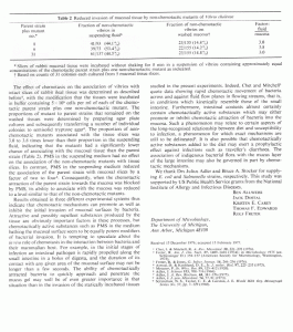
In mucosal slice adhesion, the small gut of the rabbit or mouse was cut into slices, then incubated with V Cholera in some flasks. After incubation the slices were rinsed and then put into some nutrient broth. They were then rather violently homogenized and plated out on petri dishes. The dishes were incubated and you could tell the degree to which the Cholera adheared to the slice from the number of colonies in the petri dishes.
You could study various factors affecting the adhesion to the slices with the above type of experiment. You could put various chemicals that made up the mucosa into the incubation flasks, and see if they inhibited adhesion (sticking). You could test different strains of bacteria, say motile (swim) and non-motile (can’t swim), and see what affect that had. You could let other types of bacteria compete with V Cholera, and see how the system worked. After all, there are many types of bacteria in the gut.
Another type of experiment, adhesion to ‘brush borders’, tested exactly how the Vibrio’s stuck to the cellular border of the mucosa. You could separate the surface of the mucosal cells by grinding them up, and then centrifuging them in a test tube. Under the microscope they look just like little scrub brushes. They are actually specialized cell membranes.
After isolating the membranes, you could them mix the brush border with some Vibrio’s. Looking under the microscope, you could then see bacteria sticking to the brush borders. By mixing various chemicals into the ‘soup’, you could see which ones inhibited the attachment of the Vibrios to the intestinal brush borders. Dr. Jones had done much of the work with brush borders before I arrived, and found that a sugar that is part of the brush border membrame, L-Fucose, was a potent inhibitor. This was strong evidence that L-Fucose was part of the ‘receptor’ (lock and key) that mediated colonization of the musosa by V Cholera. The only thing was, in the slice experiment, L-Fucose DID NOT inhibit adhesion. There had to be another mechanism at work, but what it was, when I joined the team, was a mystery.
Since I was a student, Dr. Freter had me begin by repeating some of the experiments that had already been done. This included the brush border experiments with L-Fucose, some of the slice inhibition experiments with various potential inhibitors, and also some histological (microscopic) studies of mouse and rabbit intestinal slices and loops that had been incubated with V Cholera. The repeat experiments came out pretty much as expected, which was good since it showed that my technique was good. But most of the other sugars I tested in the brush border and slice experiments we disappointing. We had expected to find different sugars to possibly inhibit in the slice ‘model’, since when we mixed purified fluid made from scrapings of the intestinal slices, it would inhibit adhesion to the slices. We thought there must be molecular blockers in the mucosal scrapings, much like the L-Fucose in the brush border experiments, that was preventing a lock and key fit of the Vibrio’s to receptors on the intestinal surface.
Things did not seem consistent, and I began to develop a philosophy that any result should be considered a valuable one. In other words, if d-Galactosamine did not inhibit in the brush border assay, one did not simply frown and put aside the ‘negative’ result. Rather, a piece of information was discovered that could be put in a system model, where d-Galactosamine was considered as part of the system that had no effect on adhesion. Thus, you would just work trying to find all the factors of the system, and eventually come up with a model based in facts, that could be expressed on computer or in mathematical equations. Thus you would build a knowledge base that would explain our understanding of the system.
It turned out that Dr. Freter was the right person to work with for someone with this view of biological knowledge. I found soon that he had written several papers, one titled for example “Interactions Among Interactions…” (Cite), which showed that he too held this view. So we were on the same track.
But Dr. Freter knew from his experience that for me at least, at this point in my career, the lab was the place to make discoveries. I had spent several days making system charts at my desk, when he approached me and said I should do that on my own time at home. He said I should be spending my time in the lab, not putting what we already knew into specific models. I listened to what he said, though I was rather angry about it. I wanted to see a real and informative model of the adhesion of V Cholera to the gut made. But Dr. Jones told me, that Freter held to the philosophy that you should generate as much in the lab as possible, rather than spending too much time thinking about the results. People like Jones were just the opposite, as have been many English researchers I have known. They believed in spending a lot of time cogitating about experimental results. As for me, I don’t know. In retrospect I think Dr. Freter might have had a feeling that there was something to found out about the V Cholera that outweighed putting it all ‘together’ yet. Nonetheless, for Dr. Freters class, which I took in my junior year, I wrote my paper on ‘Systems Analysis of the Adhesion of V Cholera to Mucosal Surfaces’. When he gave it back to me he told me it was the future of biology. I was pleased.
I started doing rather extensive microscopic work on the surface of the gut as colonized by the Vibrio. We wanted to get an idea of where the Vibrio’s were sticking, and how quickly they did it. We were also very interested in the mucus covering of the gut surface, and wanted to know more about it. I developed techniques that preserved the mucus, and even observed it in live, in anesthetized rabbits to see just what it’s consistency was. I found that the best results, when it comes to seeing the mucus, was to do as little to the microscopic preparation as possible. We also found that the mucus was a thin, ropy covering over the actual surface of the intestine, and could be penetrated by the vibrios, as well as inert carbon particles, quite quickly. Furthermore, most of the vibrios on the slices seemed to become embedded in the mucus covering.
But what was inhibiting the adhesion to the slices that was different than that in the brush border experiment? Brush border adheasion was inhibited by L-Fucose, but slice adhesion was not. I often stayed late in the lab, and one late afternoon I was approached by Dr. Freter as I sat at my desk. Dr. Freter asked if I knew what ‘Chemotaxis’ was. I said yes, it is the attraction of certain bacteria to a chemical agent, and even conveyed that I had read an article about it in Scientific American a few weeks before. Then Dr. Freter offered, ‘what if the thing that is inhibiting the adhesion to the (intestinal) slices is chemotaxis. I mean, maybe when we put the intestinal scrapings in with the slices and vibrios, it is negating the attraction of the vibrios to the slices by some attractive agent.’
It wasn’t my original idea, but I had what is called the ‘Ah Ha!’ reaction. I could immediately see the beauty of it. The surface of the intestinal slices emitted chemicals which attracted the vibrios. The chemicals were most concentrated at the surface of the slices. This attracted the bacteria to the slice where they stuck. If you put prepared surface scrapings in the solution that held the slices and bacteria during the experimental incubation, the gradient would be eliminated. Thus, there would be an inhibition of slice adhesion by the vibrios. We agreed on the spot that investigating the hypothesis would have first priority. I went home and told my roommate that I may be involved in a substantial discovery.
There were three major hypotheses that had to be investigated in order to discover whether chemotaxis was a mechanism that mediated the association of Cholera Vibrios with the surface of the gut. First, were chemicals that were emitted by the surface of the small intestine attractants to the cholera vibrio. Second, were the intestinal scrapings that inhibited adhesion of vibrios in the slice experiment likewise chemotactic. Finally, what was the action of chemotactic agents in the brush border model.
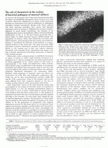
I had to devise an assay that would measure chemotaxis in V. Cholera. I found some scientific articles from other who had measured the chemical attraction of bacteria that described the physical setup of the system they used to measure the phenomenon. See Fig 1. While the setup looked reasonable, in practice it was very clumsy and inefficient. I was somewhat stuck on how to go about setting up the experiments, and was somewhat stalled. Dr. Freter saw this and said that I must get the assay to work. After some discussion we decided that we had to somehow dip capillary tubes filled with various putative attractants into small petri dishes holding vibrios in liquid broth. The result is shown in Fig 2.
The capillary assay, as it came to be called, worked very well. It was very efficient, and easy to set up. Soon we had measured the attractant qualities of the intestinal scrapings, various molecules such as L-Fucose, and ‘fractions’ of intestinal scrapings that inhibited adhesion to slices in varying degrees.
The capillary assay was rather elegant, and could be used to model the slice experiments in a very effective manner. For example, you could put vibrios in the petri dish in a solution that was basically water. In the capillary tube you could put a solution of the intestinal scrapings. This would simulate the slice experiment, with the capillary tube being the slice, and petri dish being the solution in which the slices were incubated. It was found that the vibrios would swim into the capillary tubes containing the intestinal scrapings to a greater degree that they did into tubes containing water. If you had scrapings in both the petri dish and capillary tube there would be no chemical gradient, so basically in this situation, there would be no attraction into the capillary tube. These experiments strongly agreed with the hypothesis that chemotaxis mediated the action of vibrio cholera in the slice experiments.
Several other results went further to suggest that chemotaxis was playing a major role in the colonization of the gut by V. cholera. We tested the various fractions of intestinal scrapings as to their chemotactic attraction for the Vibrios. Then we tested the degree to which these fractions inhibited colonization of slices in the slice experiment. The results were a mirror image. Those fractions that inhibited colonization to a greater extent, attracted the Vibrio’s into the capillary tubes to a greater extent.
It was found that L-Fucose, which inhibited attachment to brush borders but not to slices, was not an attractant in the capillary test. This was consistent with the working hypothesis that chemotaxis was the mechanism in the slice test, but not in the brush border assay. It was interesting that this ‘negative’ result served to support our newfound view of the system of colonization of the gut by V.Cholera, as well as my personal belief that all factors of the system studied contributed to our knowledge, not just those providing ‘positive’ results.
More tests with non chemotactic mutants of V Cholera, non-motile strains , and even microscopic observations showing ‘bands’ of bacteria forming around pieces of gut put the seal on the hypothesis that Chemotaxes is a major factor mediating attachment of V. Cholera to the intestinal wall.
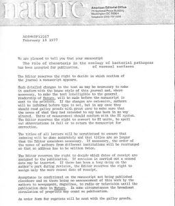
It took only about a year to prove it, and Dr. Freter sent a manuscript to Nature describing our efforts. We waited in anticipation to see if our discovery merited inclusion in the prestigious magazine. I knew that if it did, it would make a dream of mine come true. Several weeks later, on February 15, 1977, I came into the lab and Dr. Freter presented a letter to me that had come from Nature’s editor. It stated that our manuscript “The Role of Chemotaxis in the ecology of bacterial pathogens of mucosal surfaces.” Had been accepted for publication! (See Figure at end of this essay)
We were all ecstatic. I almost couldn’t believe it. In a career that can go for years with no major findings, I was the principle collaborator in a significant discovery after only 2 years. My dream had come true!
I went home to my apartment with a copy of the letter. My roommate, Mark Kandel, with whom I had attended school since Jr. High, was already there. I told him the unbelievable news. He congratulated me graciously. Then he told me he had won a Hopwood Award! He told me he had entered the competition, given by the University of Michigan school of Literature Science and the Arts with no real expectations if winning anything, but he did! And here, one of the same awards Arthur Miller had won in the 40‘s when he was a student and U-M.
So there you go. Lightening struck twice, in the same place!
What I hope to show in this essay is two fold. First, I wanted to convey to you the type of effort that goes into scientific discovery. It is a painstaking, not necessarily glorious, process that is driven by curiosity and a passion in science. Jokingly awards and discoveries may be hinted , but it is mainly a love for science. Secondly, I want to show the role that not only work, but yes indeed chance, takes place in any one individuals success or fame that may result. At least back in those days I was considered a rather bright individual by my colleagues. But what if I had not met Dr. Freter by chance when I was looking for a research position? What if things had been at a different stage in his lab’s progress? What was it exactly that made Dr. Freter suspect chemotaxis?
To be sure, opportunity favors the prepared mind. But this essay also shows that luck also can play a significant role in making your dream come true. And if it doesn’t happen, it doesn’t make you less of a person. You should know that it CAN happen to you, and you should also not be too hard on those who make an effort, but have not, or may never reach that point of success! Furthermore, it doesn’t hurt to live for the dream. It can really help you keep going in the tough times. It has helped me. So with some personal experience, I hope you have one and that your dream comes true too!
Here are the citations for the Nature Article, and another that discusses the research we did on Chemotaxis (or you may email me for a reprint at ballweiss55@gmail.com, or enter the keywords Ben Allweiss Chemotaxis in GOOGLE to access other articles on the web).
West Bloomfield, MI
October 2005
Scottsdale, AZ
Revised
December 2013